neurorradiología

28 may 2010

Intoxicación por metanol

Intoxicación por metanol
Aquí podéis ver las lesiones cerebrales de un paciente intoxicado con metanol. Las manifestaciones clínicas en urgencias fueron debilidad, visión borrosa, midriasis bilateral arreactiva y disminución progresiva del nivel de conciencia. El TAC y la resonancia mostraron necrosis hemorrágica putaminal y lesiones en sustancia blanca subcortical con realce periférico. Sólo se observó mejoría parcial durante el seguimiento del paciente. Podéis consultar el trabajo íntegro gratuitamente a texto completo en AJNR: CT and MRI findings in methanol intoxication. Blanco M, Casado R, Vázquez F, Pumar JM. AJNR. 27 (2): 452. (2006).


We present the CT and MRI findings in acute methanol intoxication in a 35 year old man who was admitted to the emergency department with weakness, blurred vision, mild bilateral areactive mydriasis and a progressive decrease in the level of consciousness. CT and MRI showed bilateral putaminal haemorragic necrosis and subcortical white matter lesions with peripheral contrast enhancement. There was only partial improvement in patient’s Glasgow Coma Scale (GCS) score during follow up.

25 may 2010

Osteopoiquilia

osteopoiquilia
La osteopoiquilia es una displasia ósea caracterizada por la presencia de múltiples lesiones osteoscleróticas. En algunas ocasiones es necesario realizar el diagnóstico diferencial con metástasis óseas escleróticas, por ejemplo de un cáncer de próstata.

18 may 2010

Mejor 1.5 Tesla

Diffusion-weighted MRI in acute stroke within the first 6 hours. 1.5 or 3.0 Tesla?

Neurology. 2010 May 12. [Epub ahead of print]
Rosso C, Drier A, Lacroix D, Mutlu G, Pires C, Lehéricy S, Samson Y, Dormont D.
From the AP-HP, Urgences Cérébro-Vasculaires (C.R., G.M., C.P., Y.S.), and AP-HP, Service de Neuroradiologie (A.D., D.L., S.L., D.D.), Université Pierre et Marie Curie, Paris VI, Hôpital Pitié-Salpêtrière, Paris, France.

Abstract

OBJECTIVES: To compare the sensitivity and specificity of 1.5-T and 3.0-T diffusion-weighted MRI (DWI) to detect hyperacute ischemic stroke lesions. METHODS: We blindly reviewed the DWI of 135 acute stroke patients and 34 controls performed at 1.5 T (n = 108) or 3.0 T (n = 61). The stroke patients all had subsequently proved carotid territory ischemic stroke and were imaged within the first 6 hours after stroke onset. Four readers (2 neuroradiologists and 2 stroke neurologists) blinded to clinical data and magnetic field strength recorded the presence of ischemic lesions on DWI and apparent diffusion coefficient (ADC) maps if necessary. Sensitivity, specificity, and false-negative rates were computed. Signal-to-noise ratio (SNR), contrast-to-noise ratio (CNR), and DWI contrasts were calculated at both field strengths. RESULTS: The accuracy of DWI in stroke diagnosis was superior at 1.5 T (98.8%) than at 3.0 T (90.9%, p = 0.03). The sensitivity decreased from 99.1% at 1.5 T to 92.5% at 3.0 T (p = 0.06) and the specificity from 97.8% to 84.1% (p = 0.002). ADC map readings did not improve accuracy, sensitivity, or specificity. The false-negative rate was 0.6% at 1.5 T and 6.1% at 3.0 T. Type of readers, stroke severity, and type of the coil did not affect diagnosis value. SNR and CNR were significantly higher at 3 T (p < 0.0001) but DWI contrast was lower (p = 0.04). CONCLUSIONS: Blind reading by 4 experts of a large series of images shows that 1.5-T diffusion-weighted MRI (DWI) is better than 3.0-T DWI for the imaging of hyperacute stroke during the therapeutic window of thrombolysis.


Vaya, vaya. Seguimos con la línea de artículos ligeramente sorprendentes, aunque no tanto. O sea que la resonancia de 3.0 T no es tan buena como el viejo cacharro de 1.5 T para el infarto hiperagudo. Espero que no empiecen a salir publicaciones de este tipo para otros tipos de exploraciones o situaciones clínicas. Aunque soy escéptico por naturaleza. Estoy seguro, no obstante, de que el mayor número de artículos defenderá los avances y progresos que con toda seguridad se van a obtener de la nueva máquina, pero se agradece una bocanada de aire fresco de gente con capacidad crítica que compense el efecto "¡ta-chan!" de la novedad tecnológica. Aprovecho para recordaros que si queréis opinar o discrepar o advertir que vuestros datos no coinciden con los de los autores del artículo, podéis hacerlo enviando un comentario.

16 may 2010

EURORAD - Radiologic Teaching Files

Esta es la fantástica base de datos europea de casos radiológicos. Rebosa cantidad y calidad. Muy aconsejable.


14 may 2010

Telangiectasia capilar

telangiectasia  capilar
Neuroimagen por resonancia magnética de un paciente con una telangiectasia capilar como hallazgo incidental.

11 may 2010

Infarto subagudo en territorio de arteria cerebral posterior derecha

infarto subagudo cerebral posterior
En la neuroimagen de hoy traemos la resonancia magnética de una paciente con un infarto subagudo en el territorio de la arteria cerebral posterior derecha. Obsérvese el realce de aspecto giriforme en secuencia T1 con contraste en el territorio de la arteria cerebral posterior derecha (abajo a la izquierda).

¿También el stent?

Carotid artery stenting compared with endarterectomy in patients with symptomatic carotid stenosis (International Carotid Stenting Study): an interim analysis of a randomised controlled trial
The Lancet, Volume 375, Issue 9719, Pages 985 - 997, 20 March 2010

Background

Stents are an alternative treatment to carotid endarterectomy for symptomatic carotid stenosis, but previous trials have not established equivalent safety and efficacy. We compared the safety of carotid artery stenting with that of carotid endarterectomy.

Methods

The International Carotid Stenting Study (ICSS) is a multicentre, international, randomised controlled trial with blinded adjudication of outcomes. Patients with recently symptomatic carotid artery stenosis were randomly assigned in a 1:1 ratio to receive carotid artery stenting or carotid endarterectomy. Randomisation was by telephone call or fax to a central computerised service and was stratified by centre with minimisation for sex, age, contralateral occlusion, and side of the randomised artery. Patients and investigators were not masked to treatment assignment. Patients were followed up by independent clinicians not directly involved in delivering the randomised treatment. The primary outcome measure of the trial is the 3-year rate of fatal or disabling stroke in any territory, which has not been analysed yet. The main outcome measure for the interim safety analysis was the 120-day rate of stroke, death, or procedural myocardial infarction. Analysis was by intention to treat (ITT). This study is registered, number ISRCTN25337470.

Findings

The trial enrolled 1713 patients (stenting group, n=855; endarterectomy group, n=858). Two patients in the stenting group and one in the endarterectomy group withdrew immediately after randomisation, and were not included in the ITT analysis. Between randomisation and 120 days, there were 34 (Kaplan-Meier estimate 4·0%) events of disabling stroke or death in the stenting group compared with 27 (3·2%) events in the endarterectomy group (hazard ratio [HR] 1·28, 95% CI 0·77—2·11). The incidence of stroke, death, or procedural myocardial infarction was 8·5% in the stenting group compared with 5·2% in the endarterectomy group (72 vs 44 events; HR 1·69, 1·16—2·45, p=0·006). Risks of any stroke (65 vs 35 events; HR 1·92, 1·27—2·89) and all-cause death (19 vs seven events; HR 2·76, 1·16—6·56) were higher in the stenting group than in the endarterectomy group. Three procedural myocardial infarctions were recorded in the stenting group, all of which were fatal, compared with four, all non-fatal, in the endarterectomy group. There was one event of cranial nerve palsy in the stenting group compared with 45 in the endarterectomy group. There were also fewer haematomas of any severity in the stenting group than in the endarterectomy group (31 vs 50 events; p=0·0197).

Interpretation

Completion of long-term follow-up is needed to establish the efficacy of carotid artery stenting compared with endarterectomy. In the meantime, carotid endarterectomy should remain the treatment of choice for patients suitable for surgery.


¿Qué está pasando con la Medicina?. Estoy confuso. Tengo la sensación de que la colocación de stents es una técnica bastante segura y eficaz. La misma impresión deben de tener cientos de profesionales que los siguen colocando. ¿Qué pasa? ¿Nos hemos dejado deslumbrar por un tratamiento nuevo y flamante una vez más? ¿Es cuestión de marketing?. Si hacemos caso a este estudio, el stenting se debe restringir a pacientes no candidatos a cirugía mientras otro estudio a más largo plazo no contradiga a éste. Definitivamente se necesita un debate más extenso, más libre, más abierto y más sano en el colectivo médico.

6 may 2010

Seguridad en Resonancia Magnética

La biblia de la seguridad en resonancia magnética. Lista completa de dispositivos médicos probados in vitro con calificación de seguros, inseguros o condicionales. Si no sabes si puedes meter o no a un paciente en la resonancia visita esta página.

Traumatismo facial

Artículo comprensible sobre fracturas maxilares y Le Fort.

4 may 2010

La vertebroplastia en entredicho

A randomized trial of vertebroplasty for osteoporotic spinal fractures.

N Engl J Med. 2009 Aug 6;361(6):569-79

Department of Radiology, Mayo Clinic, 200 First St. SW, Rochester, MN 55905, USA.

Abstract

BACKGROUND: Vertebroplasty is commonly used to treat painful, osteoporotic vertebral compression fractures. METHODS: In this multicenter trial, we randomly assigned 131 patients who had one to three painful osteoporotic vertebral compression fractures to undergo either vertebroplasty or a simulated procedure without cement (control group). The primary outcomes were scores on the modified Roland-Morris Disability Questionnaire (RDQ) (on a scale of 0 to 23, with higher scores indicating greater disability) and patients' ratings of average pain intensity during the preceding 24 hours at 1 month (on a scale of 0 to 10, with higher scores indicating more severe pain). Patients were allowed to cross over to the other study group after 1 month. RESULTS: All patients underwent the assigned intervention (68 vertebroplasties and 63 simulated procedures). The baseline characteristics were similar in the two groups. At 1 month, there was no significant difference between the vertebroplasty group and the control group in either the RDQ score (difference, 0.7; 95% confidence interval [CI], -1.3 to 2.8; P=0.49) or the pain rating (difference, 0.7; 95% CI, -0.3 to 1.7; P=0.19). Both groups had immediate improvement in disability and pain scores after the intervention. Although the two groups did not differ significantly on any secondary outcome measure at 1 month, there was a trend toward a higher rate of clinically meaningful improvement in pain (a 30% decrease from baseline) in the vertebroplasty group (64% vs. 48%, P=0.06). At 3 months, there was a higher crossover rate in the control group than in the vertebroplasty group (43% vs. 12%, P<0.001). There was one serious adverse event in each group. CONCLUSIONS: Improvements in pain and pain-related disability associated with osteoporotic compression fractures in patients treated with vertebroplasty were similar to the improvements in a control group.

Otro artículo polémico para animar a que hagáis comentarios. Este es uno de los más revolucionarios. A ver qué os parece

3 may 2010

Aracnoiditis lumbar


aracnoiditis

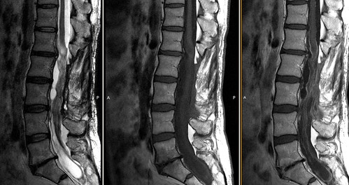
Neuroimagen por resonancia de columna lumbar, secuencias T2, T1 y T1 con gadolinio en plano sagital. El paciente acude por lumbociatalgia tras exéresis de ependimoma mixopapilar lumbar.
Se observan cambios postquirúrgicos y alteración de la disposición normal de las raíces de la cola de caballo en el saco tecal lumbar, con presencia de imágenes lineales hipercaptantes y zonas de aspecto quístico intrarraquídeo. Los hallazgos son característicos de la aracnoiditis adhesiva postquirúrgica.

¿Mejor sin filtro?

Filter-protected versus unprotected carotid artery stenting: a randomised trial.

Macdonald S, Evans DH, Griffiths PD, McKevitt FM, Venables GS, Cleveland TJ, Gaines PA.
Vascular Radiology, Freeman Hospital, Newcastle upon Tyne, UK

Cerebrovasc Dis. 2010 Feb;29(3):282-9. Epub 2010 Jan 15.

BACKGROUND: Our aim was to determine whether filter protection reduces embolisation to the brain during carotid artery stenting (CAS). METHODS: Thirty patients with symptomatic carotid artery stenosis > or =70% (North American Symptomatic Carotid Endarterectomy Trial) were randomly assigned to filter-protected or unprotected CAS. Diffusion-weighted magnetic resonance imaging (DWI) of the brain was performed before and at 3 time points after CAS. In a subset of patients, high-intensity transient signals on transcranial Doppler (TCD) were recorded with categorisation of emboli. Data were independently reviewed off-site. RESULTS: There were no significant differences in mean age, proportion of octogenarians or presenting symptoms between the groups. On procedural DWI (1-3 and 24 h after stenting), there were 7/24 (29%) and 4/22 (18%) new lesions in protected and unprotected patients respectively (p = 0.38). At 30 days there were 9/33 (26%) and 4/33 (12%) lesions in protected and unprotected patients, respectively (p = 0.1). On TCD there were significantly more signals in total as well as particulate emboli during filter-protected CAS (426.5 and 251.3) than during unprotected CAS (165.2 and 92) - p = 0.01 and 0.03, respectively. CONCLUSIONS: Filter-protected CAS is associated with an increase in new lesions on DWI and significantly higher rates of total and particulate microembolisation on TCD than unprotected CAS. The clinical significance of these findings requires further study. Copyright 2010 S. Karger AG, Basel.


Seguimos con la serie de artículos polémicos o curiosos. Si en el filtro se encuentran restos de placa (y se encuentran) ¿cómo se explica esto?

¿Embolización o clipaje? II

International Subarachnoid Trial in the Long Run: Critical Evaluation of the Long-term Follow-up Data From the ISAT Trial of Clipping Vs Coiling for Ruptured Intracranial Aneurysms.

Neurosurgery. 2010 Apr 23. [Epub ahead of print]

Raper DM, Allan R.
Northern Clinical School, University of Sydney, Royal North Shore Hospital, Sydney, Australia (Raper) Department of Neurosurgery, Royal Prince Alfred Hospital, Sydney, Australia (Allan).

The International Subarachnoid Trial (ISAT), the largest prospective randomized study into endovascular and neurosurgical treatment of ruptured intracranial aneurysms, recently reported long-term follow-up in The Lancet Neurology. In this cohort, the risk of death at 5 years was significantly lower in the coiled group, but the proportion of survivors who were independent was not statistically different between the groups, and rebleeding was higher in the coiled group. This article critically evaluates the long-term ISAT data from an evidence-based perspective and places it in the context of the overall approach to treatment of ruptured intracranial aneurysms. ISAT has been a strong driver of change in the management of ruptured aneurysms. Nevertheless, the evidence for the superiority in coiling in the long term should not be assumed from ISAT data alone. Potential biases of patient characteristics and national referral patterns, as well as the methodological problems already described from the original trial, contribute to the difficulty in interpreting differences in long-term outcomes. These new data should be regarded as Level 2b evidence, suitable for treatment recommendations but not guidelines.